Cranial Cruciate Ligament (CCL) rupture
Cranial cruciate ligament (CCL) rupture is one of the most frequent causes of hindlimb lameness in canine orthopedic patients.
Its etiology is multifactorial, involving a combination of biomechanical, conformational, degenerative, and genetic factors.
Epidemiology:
Although certain breeds show a higher predisposition, this condition can affect dogs of any size, age, or sex.
Among the most predisposed breeds are the Labrador Retriever, Rottweiler, Newfoundland, Golden Retriever, and German Shepherd.
Some smaller breeds such as the West Highland White Terrier and Yorkshire Terrier also show increased incidence, suggesting that joint conformation and biomechanics play an important role.
Clinical signs:
Patients affected by CCL disease often present with varying degrees of lameness, pain or discomfort on stifle palpation and manipulation, and joint swelling. The progression from partial to complete ligament rupture can further exacerbate instability and pain, making early recognition essential for optimal management.
Diagnosis:
Diagnosing cranial cruciate ligament (CCL) rupture in dogs is obtained by a combination of clinical orthopedic examination and diagnostic imaging.
Clinical orthopedic evaluation is the first step - Patient affected by CCL disease typically present with hindlimb lameness that may be acute or chronic. On orthopedic examination, key findings include joint effusion, pain on stifle palpation, and reduced range of motion. The main diagnostic tools are the cranial drawer test and the tibial compression test, which assess abnormal cranial translation of the tibia relative to the femur. A positive result strongly supports CCL incompetence, although these tests may require sedation in tense or painful patients.
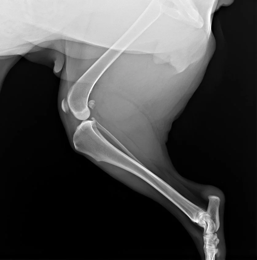
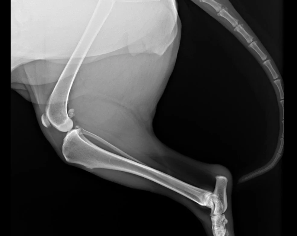
Diagnostic imaging is essential. Even though radiographs do not directly visualize the ligament, it reveals key secondary changes associated with joint instability.
Main radiographic features associated with CCL disease include joint effusion, osteophyte formation, cranial translation of the proximal tibia, and caudal displacement of the popliteal fascia. In chronic cases, degenerative changes become more evident, with osteophytes commonly observed at the caudal pole of the patella, along the femoral trochlea, and on the tibial plateau surface.
For further diagnostic imaging findings in CCL rupture: explore the clinical cases section —- > click here
Treatment:
Choosing the appropriate treatment of cranial cruciate ligament (CCL) rupture in dogs depends on several factors, including the patient’s size, age, activity level, degree of instability, and owner’s compliance and financial restraints. Treatment options can be divided into conservative (non-surgical) and surgical approaches.
Conservativemanagement may be appropriate for small dogs, sedentary patients, or cases where surgery is not feasible. This approach focuses on weight management, strict activity restriction, controlled physiotherapy, and pain management. While some dogs can achieve acceptable function, especially those under 10–15 kg, persistent instability often leads to progressive osteoarthritis.
Surgical treatment is considered the standard of care because it addresses joint instability more effectively and improves long-term outcomes.
The most commonly performed surgical techniques include:
· Tibial Plateau Leveling Osteotomy (TPLO): The tibial plateau leveling osteotomy (TPLO) is a well-described surgical procedure that treats CCL ruptures. This procedure alters the biomechanics of the stifle by leveling the tibial plateau, neutralizing cranial tibial thrust during weight bearing. This technique is widely used.
Tibial Tuberosity Advancement (TTA): This technique advances the tibial tuberosity to achieve a similar biomechanical effect, reducing the need for an intact CCL.
Extracapsular stabilization: represents a less invasive option during which a synthetic suture is placed outside the joint to simulate ligament function. It is more commonly recommended for smaller dogs or less active patients.
An adequate postoperative management plays a major role. This includes controlled and gradual return to activity, rehabilitation, pain management and weight control. Physical therapy can significantly improve limb function and recovery time.
For further indications regarding post-operative home care challenges: ——— > click here
Prognosis:
Even with appropriate surgical treatment, some degree of osteoarthritis progression is expected over time, but early and appropriate intervention can greatly improve joint stability, patient’s comfort and overall quality of life.
Summary:
Overall, the CCL disease is caused by a combination of breed-related anatomical factors (such as tibial plateau angle and ligament structure), body weight, and chronic degenerative changes rather than a single causative factor.
The progression from partial to complete ligament rupture can further exacerbate instability and pain, making early recognition essential for optimal management.