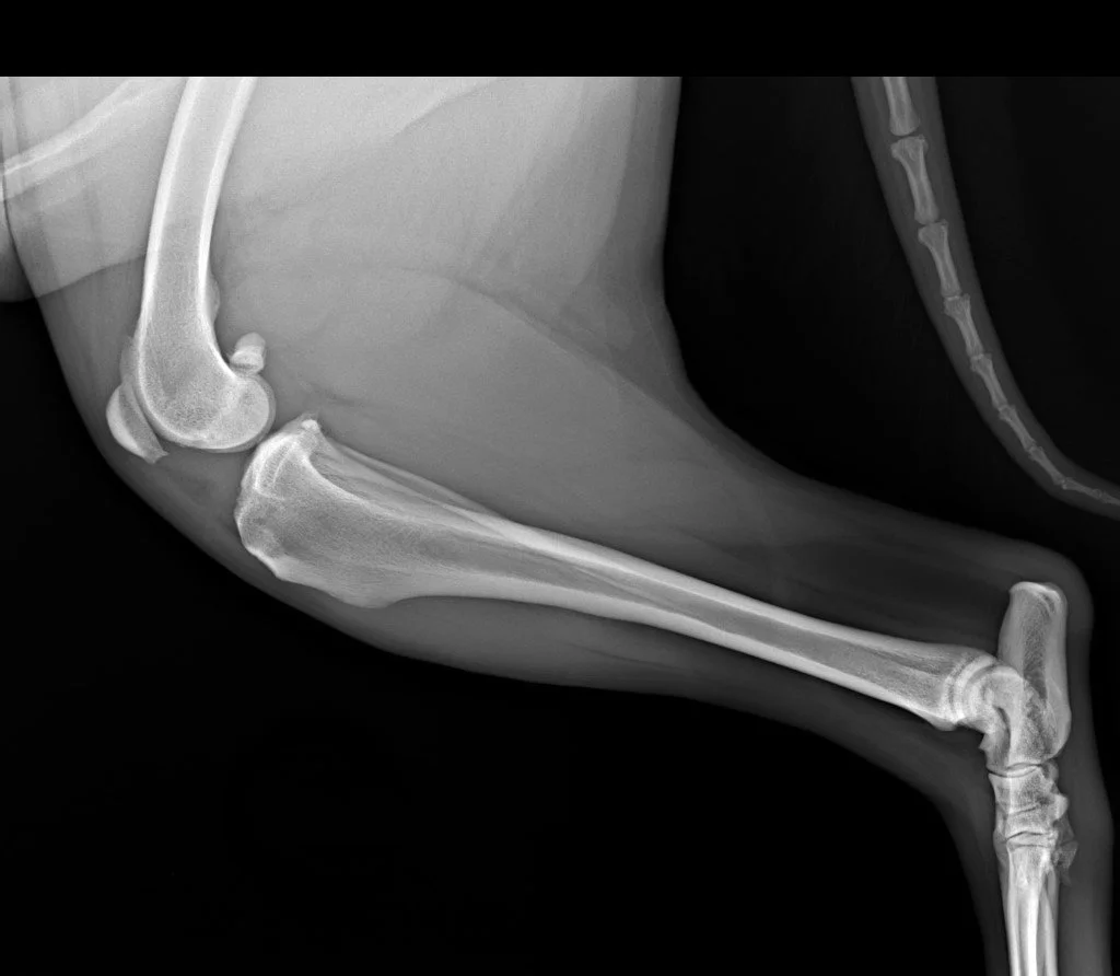
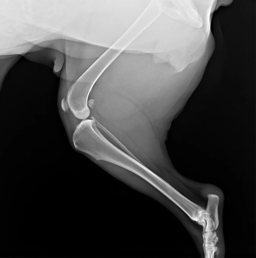
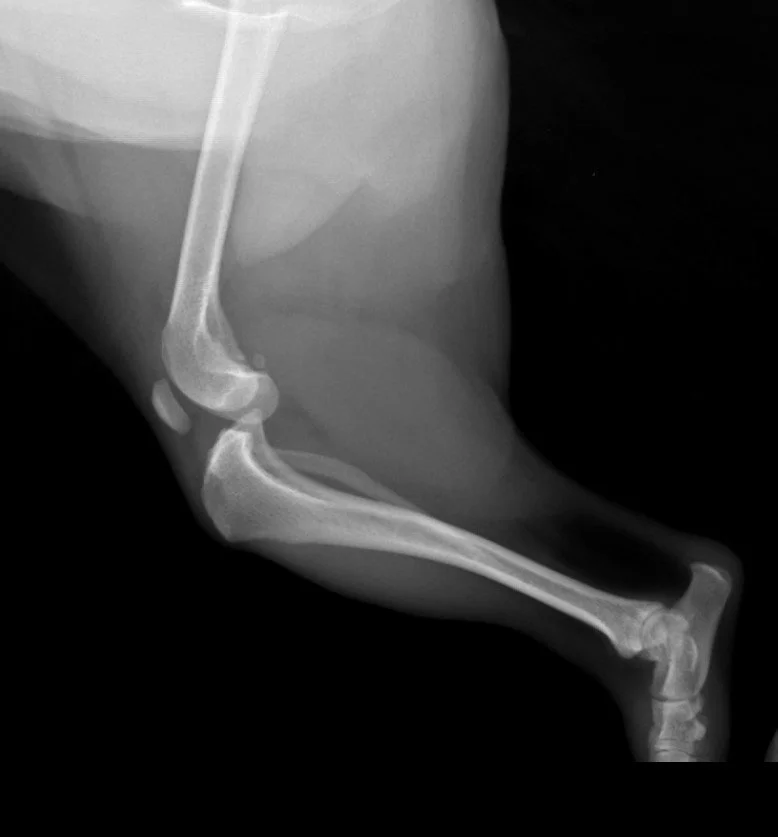
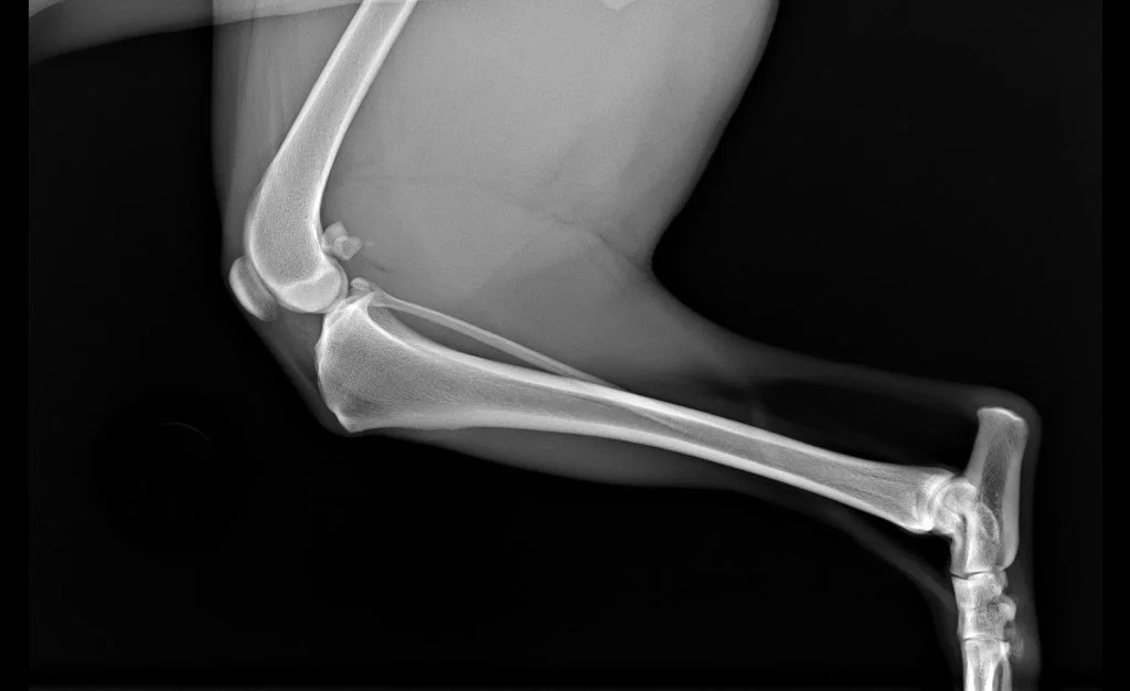
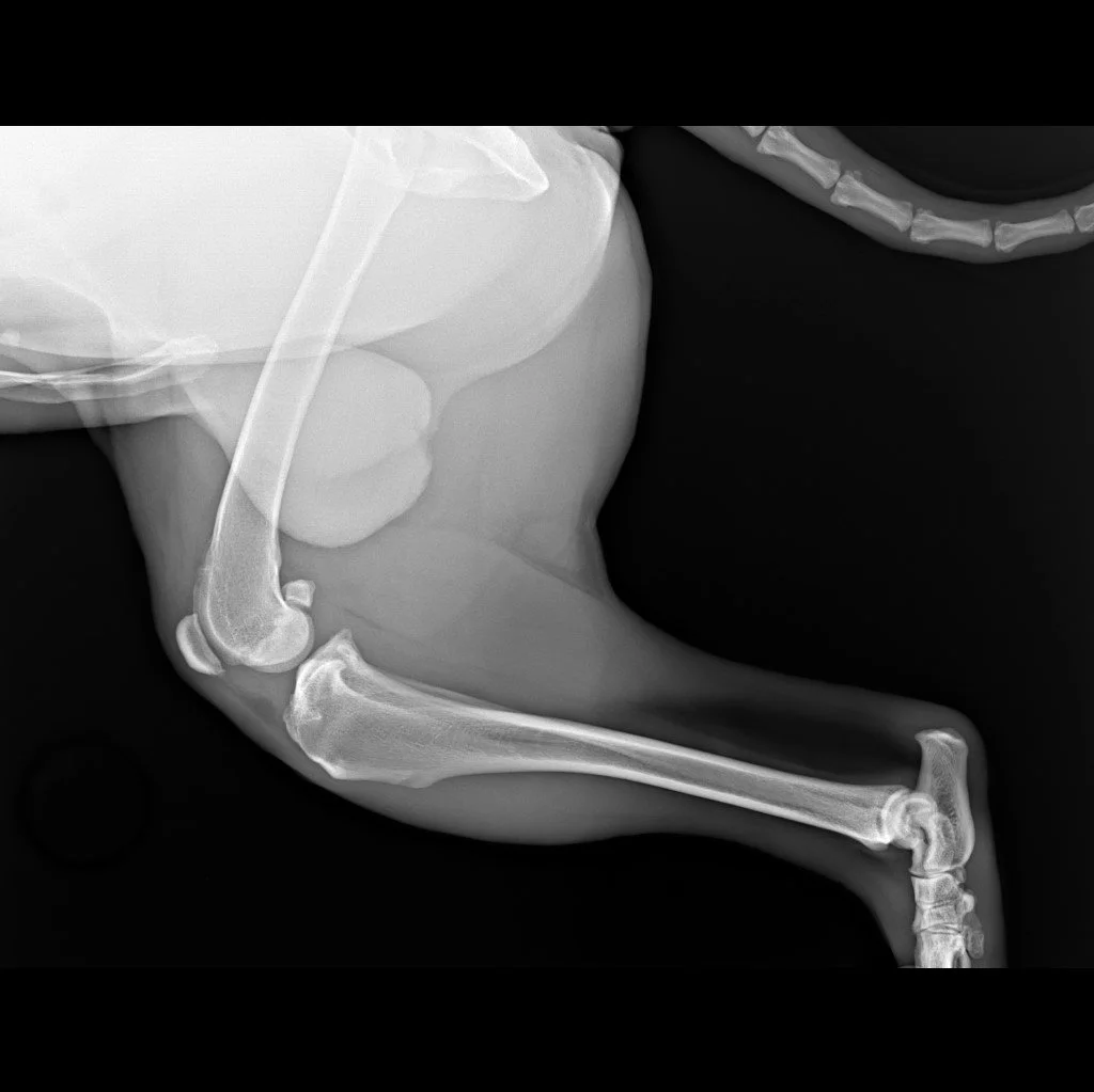
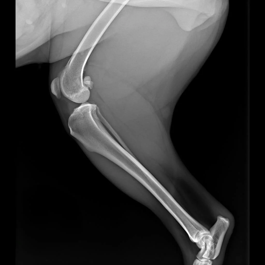
Radiographic findings in Cranial Cruciate Ligament rupture
Cranial cruciate ligament (CCL) rupture is one of the most frequent causes of hindlimb lameness in canine orthopedic patients.
Etiology is multifactorial and while certain breeds show a higher predisposition, this condition can affect dogs of any size, age, or sex.
Patients often present with varying degrees of lameness, pain on stifle palpation and joint swelling.
In this case, I share some representative radiographic finding associated with CCL disease: key findings include joint effusion, osteophyte formation, cranial translation of the proximal tibia, and caudal displacement of the popliteal fascia. In more chronic presentations, degenerative changes become more evident, with osteophytes commonly observed at the caudal pole of the patella, along the femoral trochlea, and on the tibial plateau surface.
The following radiographs illustrate these key changes, providing insight into both acute and chronic joint changes.